Departments
The analyzer is a collection of neurons, which is often called the sensory system. Any analyzer has three sections:
- peripheral - sensitive nerve endings (receptors) that are part of the sense organs (vision, hearing, taste, touch);
- conductive - nerve fibers, a chain of different types of neurons that conduct a signal (nerve impulse) from receptors to the central nervous system;
- central - a section of the cerebral cortex that analyzes and converts the signal into sensation.
Rice.
1. Analyzer departments. Each specific analyzer corresponds to a specific area of the cerebral cortex, which is called the cortical nucleus of the analyzer.
Functions
The following processes are carried out in turn in the analyzers:
- signal detection;
- signal discrimination;
- transmission and conversion of signals;
- signal recognition;
- pattern recognition.
The purpose of the transmission and transformation processes is to convey information to the brain in a convenient form. Therefore, only important information is selected, unnecessary information is eliminated.
Pattern recognition is the final operation of the analyzer. A person recognizes an image, assigns it to a category, considers it important or unimportant.
Kinds
Receptors, and therefore analyzers, can be of two types:
- external (exteroceptors) - located near or on the surface of the body and perceive environmental stimuli (light, heat, humidity);
- internal (interoceptors) - located in the walls of internal organs and perceive stimuli from the internal environment.
Rice.
2. Location of perception centers in the brain. Six types of external perception are described in the table “Human Analyzers”.
| Analyzer | Receptors | Pathways | Central departments |
| Visual | Retinal photoreceptors | Optic nerve | Occipital lobe of the cerebral cortex |
| Auditory | Hair cells of the spiral (or Corti) organ of the cochlea | Auditory nerve | Superior gyrus of the temporal lobe |
| Flavoring | Tongue receptors | Glossopharyngeal nerve | Anterior temporal lobe |
| Tactile | Receptor cells: – on bare skin – Meissner corpuscles, located in the papillary layer of the skin; – on the hair surface – hair follicle receptors; – vibrations – Pacinian corpuscles | Musculoskeletal nerves, spinal cord, medulla oblongata, diencephalon | Posterior central gyrus of the parietal lobe |
| Olfactory | Receptors of the nasal cavity | Olfactory nerve | Anterior temporal lobe |
| Temperature | Heat (Ruffini corpuscles) and cold (Krause flasks) receptors | Myelinated (cold) and unmyelinated (warm) fibers | Posterior central gyrus of the parietal lobe |
Rice.
3. Location of receptors in the skin. Internal receptors include pressure receptors, vestibular apparatus, kinesthetic or motor analyzers.
TOP 4 articles
who are reading along with this
Taste
Visual analyzer
Smell
Structure and functions of the eye
Monomodal receptors perceive one type of irritation, bimodal - two types, polymodal - several types. For example, monomodal photoreceptors perceive only light, tactile bimodal ones perceive pain and heat. The vast majority of pain receptors (nociceptors) are polymodal.
Visual analyzer.
The organ of vision is the most important of the senses, providing a person with up to 90% of information. The peripheral part of the analyzer - the organ of vision consists of the eyeball and auxiliary organs: eyelids, eyelashes, lacrimal glands, extraocular muscles (Fig. 235).
Rice. 235. Diagram of the structure of the eyeball: 1 - cornea; 2 - sclera; 3 - choroid; 4 - retina; 5 - anterior chamber of the eye; 6 - iris; 7 - posterior chamber of the eye; 8 - ciliary muscle; 9 - ligaments of Zinn; 10 - lens; 11 - vitreous body; 12 - blind spot; 13 - optic nerve; 14 - conjunctiva.
The wall of the eyeball consists of three membranes. The outer - white membrane (sclera) in the front of the eye is transparent, this part of it is called the cornea. Underneath the tunica albuginea is the choroid, which supplies the eye. In the anterior part, the choroid passes into the iris, which has a hole in the center - the pupil. The annular and radial muscles of the iris reflexively change the diameter of the pupil, regulating the amount of light entering the eye. The color of the eyes depends on the pigment of the iris. Next to the iris is the ciliary body, a muscle with the help of which the curvature of the lens changes, accommodation is carried out, adaptation to clear vision of objects located at different distances from the eye.
Between the cornea and the iris there is a cavity filled with moisture - the anterior chamber of the eye. The cavity between the iris and the lens is called the posterior chamber of the eye.
The third shell of the eyeball is the retina (Fig. 236). It contains light-sensitive cells - visual receptors, about 130 million rods, which provide black and white vision, and about 7 million cones, which provide information about color.
The maximum number of cones is located in the retina on the optical axis of the eye, opposite the pupil, this area is called the macula. In the place where the optic nerve leaves the eyeball, there are no receptors in the retina - a blind spot. The maximum number of rods is located on the periphery of the eye. The rods contain the visual pigment rhodopsin; a small amount of light is enough to decompose it. In cones, under the influence of light, iodopsin decomposes, but more light is needed to excite the cones.
The retina consists of several layers of cells: the outer one, adjacent to the choroid, is a layer of black pigment cells. This layer absorbs light, preventing its scattering and reflection. Then there is a layer containing rods and cones, followed by three more layers of cells.
Rice. 236. Structure of the retina: 1 - pigment cells; 2 - cones; 3 - sticks; 4 - bipolar cells; 5 - ganglion cells; 6 - amacrine cells.
The vitreous fills the entire cavity of the eye and is formed by a transparent gelatinous substance. Between the vitreous body and the posterior chamber of the eye is the lens, an elastic transparent formation in the form of a biconvex lens. The lens refracts light rays and brings them into focus on the retina. With the help of the ligaments of Zinn, it is attached to the ciliary (ciliary) muscle. Light passes through the cornea, the fluid in the anterior chamber of the eye, enters the posterior chamber through the pupil, passes through the lens, the vitreous body, and reaches the retina. Light rays undergo the greatest refraction in the cornea and lens, the image on the retina is reduced and inverted.
Accommodation is carried out due to the contraction of the ciliary muscle, while the ligaments of Zinn relax and the lens, due to its natural elasticity, becomes more convex. The ciliary muscle rests when a person looks into the distance, while the ligaments of Zinn are stretched and the lens flattens (Fig. 237).
Rice. 237. Change in the curvature of the lens: the ciliary muscle is relaxed above, contracted below.
With age, the lens often loses its elasticity and becomes flatter, the image from nearby objects goes behind the retina - senile farsightedness develops.
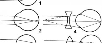
With congenital myopia, the eyeball is elongated, the focal length is closer to the retina and the image from distant objects is not sharp; with congenital farsightedness, the eyeball is shortened and the focal length goes behind the retina (Fig. 239).
Rice. 239. Refraction in normal (1), myopic (2) and farsighted eyes and optical correction of myopia (4) and farsightedness (5).
Nerve impulses travel along the fibers of the optic nerve to the posterior parts of the occipital lobes, and axons from the left halves of the retina of both eyes are sent to the left hemisphere, and from the right halves to the right. In this case, the axons from the medial halves intersect, forming a optic chiasm (Fig. 238).
Rice. 238. Diagram of human visual pathways: 1 - optic nerve; 2 - visual chiasm; 3 - geniculate bodies; 4 - visual cortex.
When the light intensity changes, a reflex change in the diameter of the pupil occurs. The sphincter muscles and constrictors are innervated by the parasympathetic nerves, the radial muscles, the dilators of the pupil, are innervated by the sympathetic nerves, so fear and pain lead to dilation of the pupils, it is not for nothing that they say: “Fear has big eyes.”
The cones in the retina are divided into three groups, some are excited by red light, others by green, and others by blue. If some group of cones does not work, diseases develop in which a non-human person can distinguish certain colors.
With a lack of vitamin A, the pigment rhodopsin in rods is not formed, and a person has difficulty seeing in the dark - the disease is called “night blindness”. Contamination of the mucous membrane of the eyelids (conjunctiva) leads to inflammatory processes - conjunctivitis. Reading in a moving vehicle, reading while lying down, and prolonged work with a computer lead to overwork of the ciliary muscle and a decrease in visual acuity. When working with small objects it should be at least 30-35 cm, the workplace should be well lit.
Characteristics
Analyzers, regardless of type, have a number of common properties:
- high sensitivity to stimuli, limited by the threshold intensity of perception (the lower the threshold, the higher the sensitivity);
- difference (differentiation) of sensitivity, allowing one to distinguish stimuli by intensity;
- adaptation, which allows you to adjust the level of sensitivity to strong stimuli;
- training, manifested both in a decrease in sensitivity and in its increase;
- preservation of perception after the cessation of the stimulus;
- interaction of different analyzers with each other, allowing you to perceive the completeness of the outside world.
An example of a peculiarity of the analyzer's operation is the smell of paint. People with a low threshold for sensitivity to odors will smell more strongly and react actively (lacrimation, nausea) than people with a high threshold. Analyzers will perceive a strong odor more intensely than other surrounding odors. Over time, the smell will not be felt sharply, as adaptation will occur. If you constantly stay in a room with paint, the sensitivity will become dull. However, when you leave the room into the fresh air, you will feel the smell of paint for some time.
Test on the topic
- /10
Question 1 of 10What is the conductor section of the analyzer?
Start test
Hall of Fame
To get here, take the test.
- Anastasia Skorinova
10/10
- Dmitry Lyutik
8/10
- Sergey Ardashev
10/10
- Alina Kozlova
10/10
- Pavel Zherebtsov
10/10